Breast cancer is a common malignant tumor. The advance of breast cancer treatments is hindered in part by challenges in translating to humans the successes achieved indistantly related rodent species. To bridge that evolutionary gap, scientists have begun to turn to an alternative model organism, the tree shrew (Tupaiabelangerichinensis). Tree shrews have increasingly become an attractive experimental animal model for human diseases, particularly for breast cancer due to spontaneous breast tumors and their close relationship to primates and by extension to humans. On the other hand, Tree shrews are superior to classical primates because tree shrew is easier to manipulate, maintain and propagate.
Recently, the research team of Professor Ceshi Chen has developed a new method for establishment of mammary tumors in tree shrew. They used a lentivirus overexpressing the PyMT oncogene to infect tree shrew mammary gland epithelial cells to induced mammary tumors. Most animals developed mammary tumors within 3 weeks of infection, and all animals exhibited tumors by 7 weeks. Among these, papillary carcinoma is the predominant tumor type. One case showed lymph node and lung metastasis. Interestingly, the expression levels of phosphorylated AKT, ERK and STAT3 were elevated in 41–68% of PyMT-induced mammary tumors, but not all tumors. Finally, we observed that the growth of PyMT-induced tree shrew mammary tumors was significantly inhibited by Cisplatin and Epidoxorubicin. PyMT-induced tree shrew mammary tumor model may be suitable for further breast cancer research and drug development, due to its high efficiency and short latency.
The main findings of this study have been published in International Journal of Cancer (http://onlinelibrary.wiley.com/doi/10.1002/ijc.29814/abstract). Due to potential applications of generating breast carcinoma tree shrew model, the research team has been granted a patent. The patent has been executed at Guangxi Medical University recently.

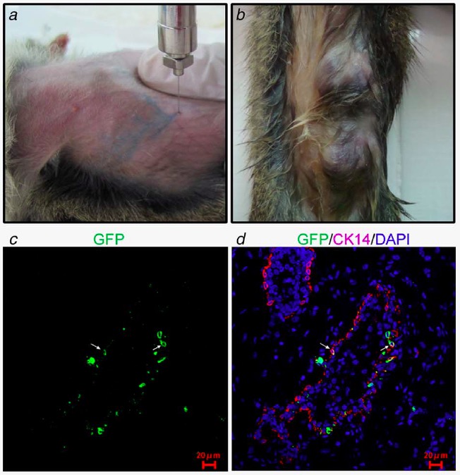
Induction of tree shrew mammary tumors using lentivirus expressing PyMT by intraductal injection. a. Lentivirus were injected into tree shrew mammary ducts and traced by trypan blue. b. Tree shrews developed mammary tumors after injecting lentivirus expressing PyMT. c, d. Immunofluorescence analysis of PyMT virus infected mammary cells. GFP as stained in green and CK14 was stained in red. Nuclear DNA was stained with DAPI (in blue). c shows GFP only and d shows a merged picture for both GFP and CK14. The white arrows indicates GFP/CK14 double positive cells.
(By Chuan-Huizi Chen)
Contact: Ceshi Chen
Email: chenc@mail.kiz.ac.cn
