Vertebrate evolution was accompanied by two rounds of whole genome duplication followed by functional divergence in terms of regulatory circuits and gene expression patterns. As a basal and slow-evolving chordate species, amphioxus is an ideal paradigm for exploring the origin and evolution of vertebrates.
A research group, led by developmental biologist Prof. MAO Bingyu from the Kunming Insitute of Zoology of the Chinese Academy of Sciences, has reported the single-nucleus RNA-seq (snRNA-seq) and single-cell assay for transposase-accessible chromatin (ATAC)-seq (scATAC-seq) atlas for amphioxus, a cephalochordate and key animal for understanding the evolution of vertebrates.
The findings were published in Cell Reports on June 21.
Transcriptomic and chromatin-accessibility profiling at the single-cell level have been increasingly used to dissect the early developmental program in model and non-model organisms, including humans, mice, zebrafish, frogs, and sea squirts.
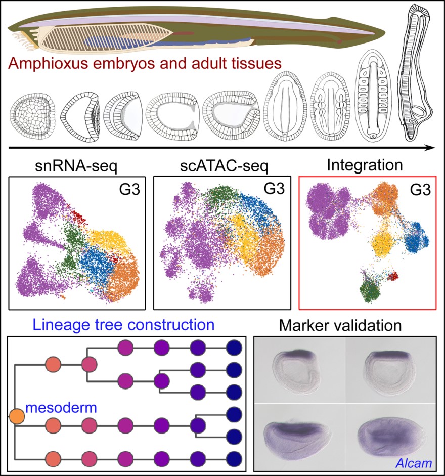
To dissect the mechanisms of cell lineage specification and the logic of the regulatory network of amphioxus embryos and shed light on the origin of vertebrates, MAO and his colleagues reported the snRNA-seq and scATAC-seq analysis for different stages of the amphioxus Branchiostoma floridae, covering embryogenesis and adult tissues.
With these datasets generated, the team built the developmental tree for amphioxus cell fate commitment and lineage specification, and characterized the underlying key regulators through integrated analysis.
In addition, cross-species analysis also revealed similar developmental trajectories, genetic regulatory networks, and regulatory modules in amphioxus and other chordate model organisms.
[Note: I don’t think it makes sense to say “other chordate models of amphioxus.” (This is how the sentence was originally worded.) Anything included in the category of “amphioxus” would already be covered by saying “. . . models in amphioxus . . .” “Chordate” is a higher (larger) category than amphioxus; it includes amphioxus. Therefore, to say “other chordate model organisms” would mean chordates that are NOT amphioxus. For this reason, saying “other chordate model organisms of amphioxus” seems to negate the author’s meaning.]
The data generated in this study are available on an online platform, AmphioxusAtlas, at https://lifeomics.shinyapps.io/shinyappmulti/.
“The data could be valuable for understanding the origin and evolution of chordates and throw light upon the genetic and epigenetic mechanism underlying the phenotype novelty of chordates during evolution.” said Prof. MAO, co-corresponding author of the study.
Results of the study, entitled “Joint profiling of gene expression and chromatin accessibility during amphioxus development at single cell resolution,” were published in Cell Reports on June 21.
This project was financially supported by grants from the Strategic Priority Research Program of the Chinese Academy of Sciences, the National Key Research and Development Program of China, and the National Natural Science Foundation of China. The first author, MA Pengcheng, was supported by the Youth Innovation Promotion Association of the Chinese Academy of Sciences.

Schematic presentation of the main findings of this research: (1) a snRNA-seq atlas of amphioxus developmental embryos and multiple adult tissues and a scATAC-seq atlas of amphioxus developmental embryos; (2) a developmental tree for amphioxus cell fate commitment and lineage specification; and (3) Computational identification and validation of lineage specific markers.
(By MA Pengcheng , Editor: YANG Yingrun )
Contact:
MAO Bingyu, Ph.D
Kunming Institute of Zoology, Chinese Academy of Sciences
Email: mao@mail.kiz.ac.cn
Tel: 0(86)871-68125418
